Prenatal diagnosis
Prenatal diagnosis
Nowadays medicine has leaped forward and allows to evaluate the health of the child is still at the stage of fetal development. This assessment is of great importance, as a percentage of intrauterine fetal malformations and inherited diseases are not declining. Prenatal diagnosis of fetal allows timely identification of virtually all deviations from the norm and to take the necessary measures.
Prenatal diagnosis: what is it?
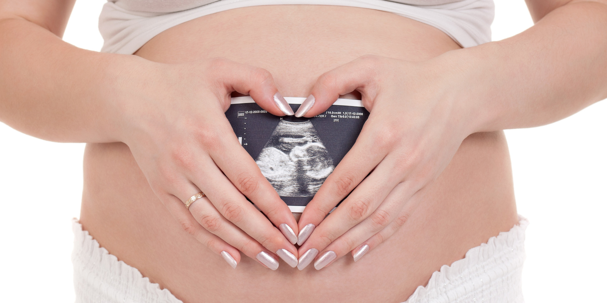
Antenatal and prenatal diagnosis – a collection of fetal research, which reveals or deny fetal malformations, chromosomal and genetic disorders of the child. Prenatal diagnosis is very young, but successfully developing branch of reproductive medicine. After receiving the results of the study carried out medical and genetic counseling of spouses and solved questions: Does it make sense to prolong pregnancy if treatment is possible with a child-diagnosed pathology in utero or after birth and how to prevent deviations from the normal development of the fetus in the next pregnancy.
Methods of prenatal diagnosis
All methods of prenatal diagnostics are divided into 2 groups. The first relates minimally invasive or non-invasive prenatal diagnosis (prenatal screening), including:
- Ultrasonography;
- Study the pedigree of parents;
- Conducting genetic research of the spouses;
- Doplerometric ultrasound (evaluation of blood flow in the system, the placenta mother fruit) on the testimony;
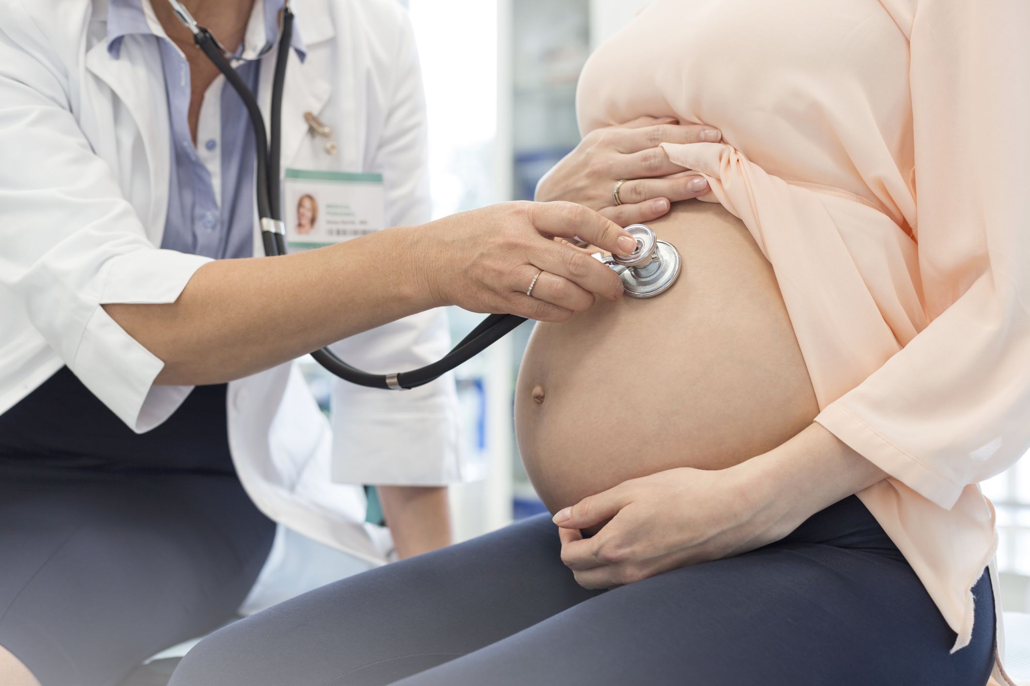
- Cardiotocography (held from 32 weeks, according to testimony from 28 weeks);
- Blood on the content of serum markers (“to deformities of the fetus”).
The second group includes invasive techniques that involve surgical penetration into the uterus:
- Chorionic villus sampling;
- Platsentotsentez;
- Cordocentesis;
- Amniocentesis;
- Biopsy of fetal tissue.
Noninvasive prenatal diagnosis
Prenatal screening (sifting or sorting) is mandatory to all pregnant women and includes 2 major studies that reveal the gross malformations and fetal pathology markers.
Ultrasonography
Ultrasonography is a safe method and should be performed during pregnancy for at least 3 times and on certain dates: 10 – 14 weeks 22 – 24 weeks and at 32 – 34 weeks. Deviation from the recommended timing greatly reduces the rate of detection of pathology. Therefore, at the first ultrasound certain signs that gross pathology before the 10th week has not yet appeared, and after 14 weeks has disappeared. However, even during the second ultrasound is not always possible to identify abnormalities and malformations (e.g., small defects in the heart septum). Therefore, ultrasound is obligatory (in any case) is complemented by research on fetal blood markers.
The methodology of the US:
Transabdominal study
It is carried out by means of transabdominal transducer that emits ultrasound waves. The sensor is found on the surface of the anterior abdominal wall, and the waves transmitted by them, are reflected by the tissues of the future baby and processed by a computer. After that, the monitor is formed sonogram – an image that is described by a doctor. Transabdominal study perform better in the second – third trimesters.
Transvaginal investigation
Is preferably carried out in the early stages of gestation. Vaginal probe placed in the condom is inserted into the vagina.
What reveals the US:
- Embryo localization (uterine or ectopic pregnancy);
- Number of fruit;
- Gestational age in weeks;
- Fetal growth retardation;
- Missed abortion;
- Sex of the child;
- Placental localization (previa, a low placentation);
- The condition of the placenta (heart attack, calcification, degree of maturity);
- The amount of amniotic fluid (oligohydramnios or multilateral);
- State cord, the number of vessels in her umbilical node;
- The tone of the myometrium (hypertonicity with the threat of interruption or premature birth);
- Fetal heart and his character (bradycardia, tachycardia);
- Impaired blood flow in the vessels of the placenta;
- Fetal abnormalities (primarily neural tube defects, heart and kidney, liver and intestinal pathology, condition of the limbs and the facial region of the skull);
- Early identification of specific symptoms of Down’s syndrome (up to 12 weeks) – the width of the neck and neck area;
- Position (longitudinal, transverse, oblique) and presentation (head, pelvic, facial) fetal.
In addition, ultrasound can diagnose molar pregnancy and anembryonic gestation (no nucleus).
Biochemical screening
For the biochemical screening of pregnant investigated venous blood taken within the period of 15 – 20 weeks (optimally 16 – 18). The first stage of the screening – the “double test” will be held at 9 – 13 weeks, these terms are defined by placental proteins PPAP-R and hCG is rare. The second step is carried out biochemical screening in the second trimester of pregnancy and is determined by the content of alpha-fetoprotein (AFP), human chorionic gonadotropin (hCG) and free estriol. The Russian Federation has studied only the first 2 marker.
What reveals biochemical screening?
- Down syndrome or trisomy;
- Abnormalities of the brain and spinal cord (no brain – anencephaly, herniation of the brain and spinal cord);
- Chromosomal abnormalities.
- The advantages of biochemical screening include:
- High-performance (detection of Down’s syndrome and neural tube defects up to 70%);
- Early diagnosis of fetal pathology (15 – 22 weeks), even when the pregnancy can be interrupted;
- No risk to the fetus.
The disadvantage is the impact of various factors (multiple pregnancy, complications of gestation, genital organs and other diseases of women) on the reliability of the results. In view of what the study of biochemical markers may show false negative or false positive result.
In suspected cases of abnormalities of biochemical markers assigned US higher level (in the perinatal center or the regional / republican hospital) and carrying out invasive prenatal diagnosis.